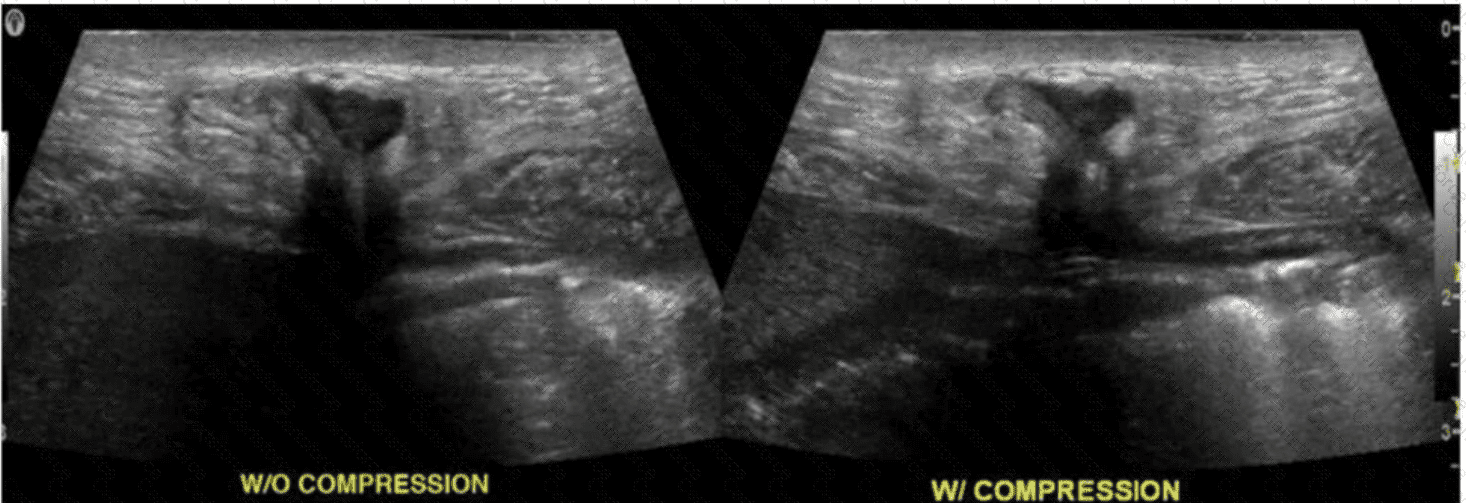
The ultrasound image demonstrates a fluid-filled structure in the posterior knee region, consistent with a Baker cyst (also called a popliteal cyst). A Baker cyst is a synovial fluid-filled sac arising from the posterior medial aspect of the knee joint, typically extending between the medial head of the gastrocnemius and the semimembranosus tendon.
The history of delayed-onset claudication (pain in the calf when walking) two weeks after this image was obtained is strongly suggestive of a ruptured Baker cyst. When a Baker cyst ruptures, synovial fluid may track inferiorly into the calf, producing pain, swelling, and clinical symptoms that mimic deep vein thrombosis (DVT) or arterial insufficiency (e.g., pseudothrombophlebitis syndrome).
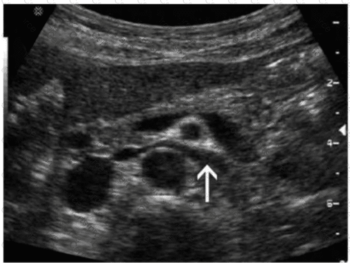
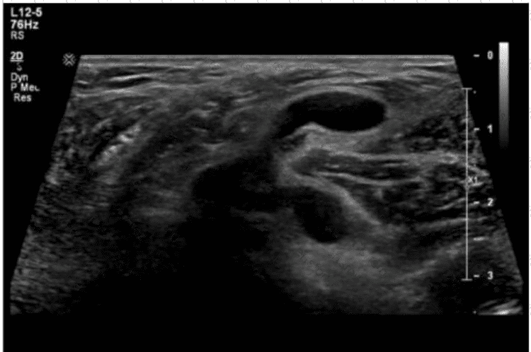
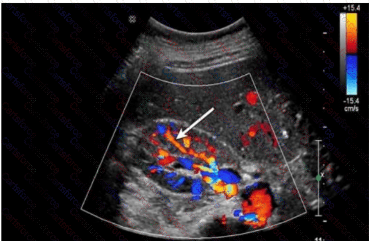
Ultrasound findings consistent with a ruptured Baker cyst:
Complex fluid collection tracking along muscle fascial planes (hypoechoic to anechoic)
Posterior calf swelling and tenderness
Absence of thrombus in the deep venous system
Crescent-shaped fluid may be seen between muscle compartments
Why the other choices are incorrect:
A. Neuropathy: Would not show fluid-filled structures on ultrasound and would not present with calf swelling.
B. Infected hematoma: May appear complex, but would require a history of trauma or anticoagulation and systemic signs (fever, redness).
C. Thrombophlebitis: Involves a thrombosed superficial vein with wall thickening and surrounding inflammation, which is not seen in this image.
[References:, American Institute of Ultrasound in Medicine (AIUM). Practice Guidelines for Musculoskeletal Ultrasound Examination, 2020., Bianchi S., Martinoli C. Ultrasound of the Musculoskeletal System. Springer, 2007. Chapter: Knee Region — Popliteal Fossa and Baker’s Cyst, pp. 433–437., Radiopaedia.org. Ruptured Baker cyst: https://radiopaedia.org/articles/ruptured-bakers-cyst, , ]