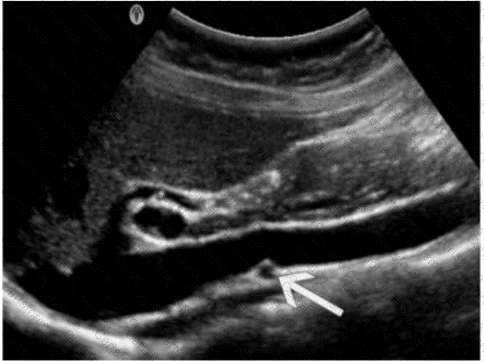
The ultrasound image demonstrates a transverse view of the abdominal vasculature, where the arrow is pointing to a circular vascular structure anterior to the aorta and posterior to the body of the pancreas — consistent with the superior mesenteric artery (SMA).
The SMA originates from the anterior aspect of the abdominal aorta just below the level of the celiac trunk and courses anterior to the left renal vein and uncinate process of the pancreas. On transverse ultrasound, it is often seen in cross-section as a round, pulsatile structure with echogenic walls, situated just anterior to the aorta. This appearance is known as the “target sign” or “bull's-eye” appearance.
Vessel Position Landmarks (transverse plane):
Aorta: Posterior and central
SMA: Just anterior to the aorta
Left renal vein: Passes between the aorta and SMA (nutcracker location)
Right renal artery: Courses posterior to the IVC toward the right kidney
Differentiation from other options:
A. Proper hepatic artery: Typically visualized within the liver hilum (portal triad), not in this anatomic location.
C. Left renal vein: Seen in transverse as a longer, oval structure crossing anterior to the aorta and posterior to the SMA.
D. Right renal artery: Arises laterally from the aorta and courses posterior to the IVC — not visualized in this axial midline location.
[References:, Rumack CM, Wilson SR, Charboneau JW, Levine D. Diagnostic Ultrasound. 5th Edition. Elsevier, 2018. Chapter: Vascular Anatomy and Abdominal Vessels, pp. 471–475., American Institute of Ultrasound in Medicine (AIUM) Practice Parameter for the Performance of an Ultrasound Examination of the Abdomen and/or Retroperitoneum, 2020., Radiopaedia.org. Superior mesenteric artery: https://radiopaedia.org/articles/superior-mesenteric-artery, —, ]
Submit