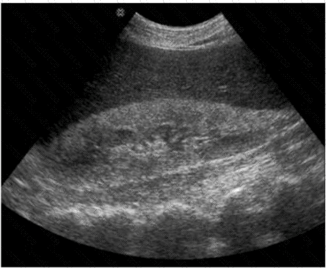
The ultrasound image demonstrates diffuse increased echogenicity of the liver parenchyma with posterior beam attenuation (acoustic shadowing), findings that are consistent with hepatic steatosis (fatty liver disease). The liver appears brighter than normal, and the vascular markings, particularly of the portal veins, are obscured due to the increased parenchymal echogenicity.
Hepatic steatosis refers to the abnormal accumulation of fat within hepatocytes and is commonly associated with obesity, diabetes, alcohol use, and metabolic syndrome.
Classic sonographic features of hepatic steatosis include:
Diffuse hyperechogenicity (“bright liver”)
Poor visualization of intrahepatic vessels and diaphragm
Posterior acoustic attenuation
Increased hepatic echogenicity relative to the renal cortex
Differentiation from other options:
A. Acute hepatitis: Usually presents with normal or slightly decreased echogenicity, "starry sky" appearance due to prominent portal triads and periportal edema.
C. Medullary sponge kidney: A renal condition with echogenic medullary pyramids, not hepatic.
D. Acute medical renal disease: Affects the kidneys, often with bilateral renal enlargement and increased cortical echogenicity—again not related to liver imaging.
[References:, Rumack CM, Wilson SR, Charboneau JW, Levine D. Diagnostic Ultrasound. 5th Edition. Elsevier, 2018. Chapter: Liver, pp. 93–97., American College of Radiology (ACR) Practice Parameter for the Performance of an Ultrasound Examination of the Abdomen and/or Retroperitoneum, 2021., Radiopaedia.org. Fatty liver (ultrasound): https://radiopaedia.org/articles/fatty-liver-ultrasound, , ]
Submit