Which scanning approach was utilized to obtain this image?
Which type of artifact is indicated by the arrows on this image?

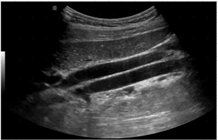
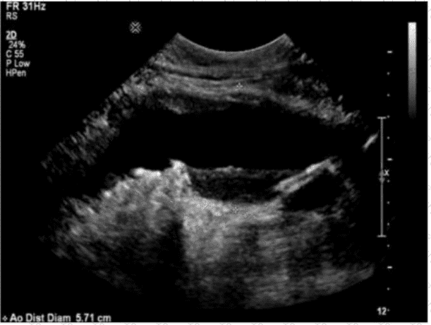
Which sonographic finding is most consistent with this image of the abdominal aorta?
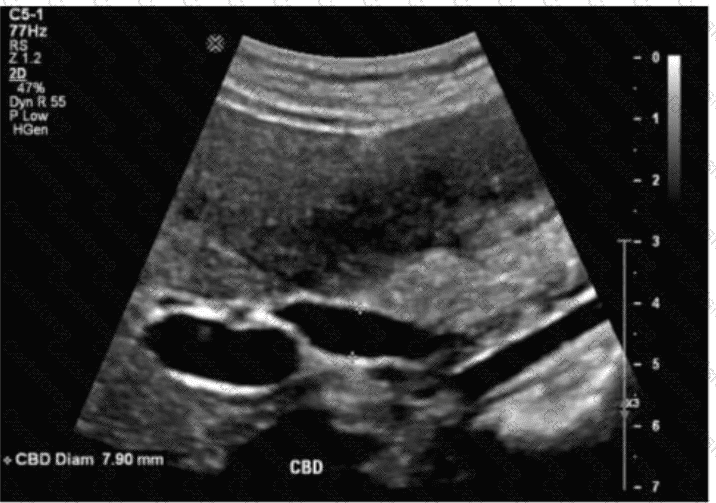
Which term best describes the common bile duct measured in this image of a postcholecystectomy patient?
Which condition puts the patient at greatest risk for a hematoma as a result of biopsy?
Which vessel is typically seen with an echogenic ring of fat when imaging the upper abdominal mesenteric circulation?
Which type of choledochal cyst is the most common?
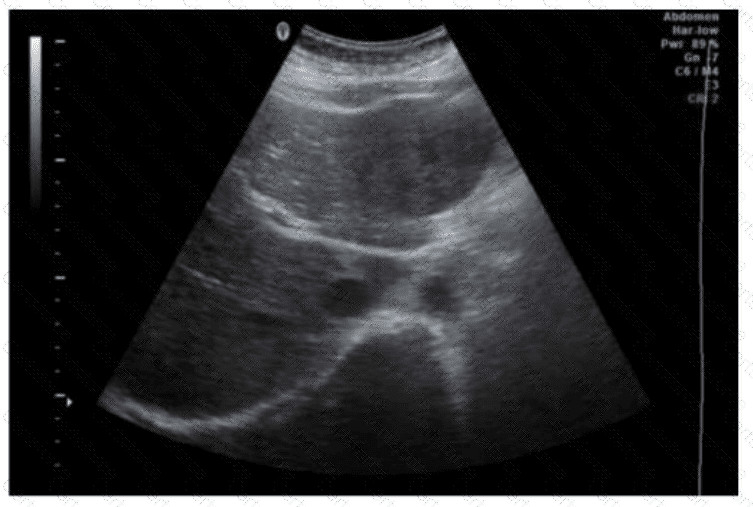
In which segment is the solid mass located in this transverse image of the liver?
Which technique would best assist the sonographer to verify the finding in this image obtained from the right upper quadrant?
Which condition is most likely to develop after splenic trauma?