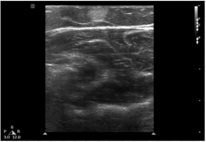
The ultrasound image demonstrates a well-defined, ovoid, hypoechoic to isoechoic mass within the subcutaneous tissue of the abdominal wall. The lesion appears compressible and shows linear striations parallel to the skin surface — a classic appearance of a lipoma.
Lipomas are the most common benign soft tissue tumors and frequently arise in the subcutaneous tissue. They are composed of mature adipose tissue and are typically asymptomatic unless large or compressing adjacent structures.
Sonographic features of a lipoma:
Isoechoic to mildly hyperechoic or hypoechoic relative to subcutaneous fat
Oval or elliptical in shape with well-defined margins
Internal linear striations or “feathered” echotexture
Compressible and non-vascular on Doppler imaging
Located in subcutaneous fat plane parallel to the skin surface
Differentiation from other options:
B. Fibroma: Typically appears as a homogeneous, hypoechoic mass but is far less common in the abdominal wall.
C. Desmoid: Appears as an ill-defined or infiltrative mass within deeper soft tissues; more heterogeneous and may distort surrounding tissue planes.
D. Metastasis: Often more irregular, heterogeneous, and may show increased vascularity or invasion into adjacent structures.
[References:, Rumack CM, Wilson SR, Charboneau JW, Levine D. Diagnostic Ultrasound. 5th Edition. Elsevier, 2018. Chapter: Musculoskeletal and Soft Tissue Ultrasound, pp. 1448–1450., American Institute of Ultrasound in Medicine (AIUM) Practice Parameter for the Performance of a Diagnostic Ultrasound Examination of Soft Tissue Structures, 2020., Radiopaedia.org. Lipoma (ultrasound): https://radiopaedia.org/articles/lipoma-ultrasound, , ]
Submit