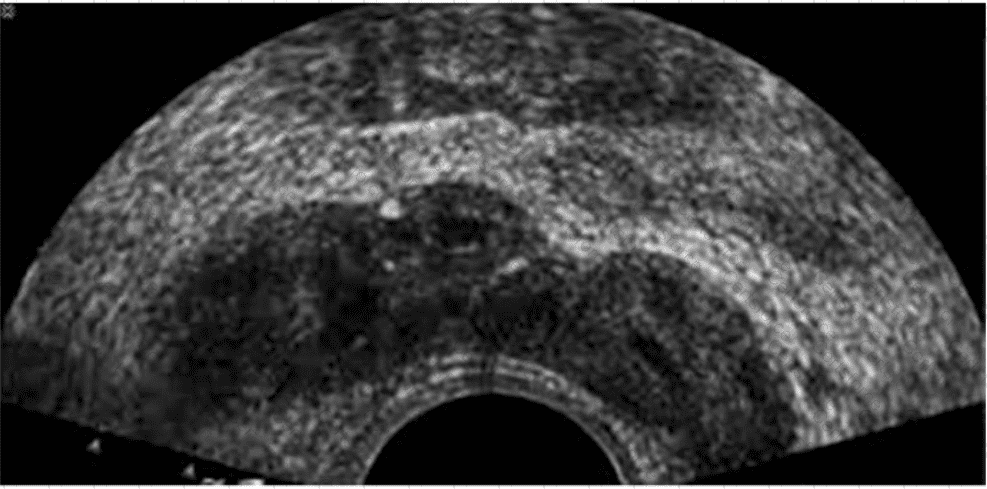
The ultrasound image shown is a transverse endorectal (transrectal) ultrasound, commonly used to evaluate the prostate and adjacent structures. The two hypoechoic (dark) oval-shaped structures seen superior and posterior to the prostate are characteristic of the seminal vesicles.
The seminal vesicles are paired, elongated glands located superior and posterior to the base of the prostate and are best visualized in transverse planes on endorectal imaging. They appear as hypoechoic or anechoic structures with internal septations, depending on the degree of fluid content.
In contrast:
The urethra appears as a central echogenic linear structure within the prostate.
The prostate base is more inferior in the scan and is visualized just above the urethra.
The ejaculatory ducts are usually not as prominently visualized and are located medial to the seminal vesicles, entering the prostate near the verumontanum.
This image most clearly demonstrates the bilateral seminal vesicles.
[References:, Rumack CM, Wilson SR, Charboneau JW, Levine D. Diagnostic Ultrasound, 5th ed. Elsevier; 2017., ACR–AIUM–SRU Practice Parameter for the Performance of an Ultrasound Examination of the Prostate (2021)., Hagen-Ansert SL. Textbook of Diagnostic Sonography, 8th ed. Elsevier; 2017., , , ]
Submit