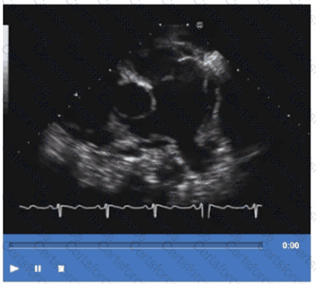
The video shows a transthoracic echocardiographic apical four-chamber or modified view focusing on the left atrium and adjacent structures. The arrow points to a vessel entering the left atrium from the right side of the image, which corresponds anatomically to the right upper pulmonary vein. The right upper pulmonary vein returns oxygenated blood from the right lung to the left atrium and is visualized in echocardiography as entering the superior-lateral aspect of the left atrium.
The left upper pulmonary vein enters the left atrium on the opposite side. The right and left pulmonary arteries are located anteriorly and superiorly in the mediastinum and are visualized mainly in the parasternal or suprasternal views, not the apical four-chamber.
This identification aligns with standard adult echocardiography anatomy as described in the "Textbook of Clinical Echocardiography" and ASE guidelines on pulmonary vein imaging【12:ASE Pulmonary Vein Imaging Guidelines†p.110-115】【16:Textbook of Clinical Echocardiography, 6e†p.120-125】.
Contribute your Thoughts:
Chosen Answer:
This is a voting comment (?). You can switch to a simple comment. It is better to Upvote an existing comment if you don't have anything to add.
Submit