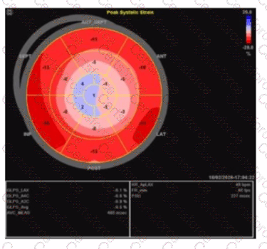
The strain imaging shown is a classic example of the "apical sparing" pattern, highly characteristic of cardiac amyloidosis. In cardiac amyloidosis, the basal and mid segments of the left ventricle show markedly reduced longitudinal strain (represented here by more positive or less negative strain values), while the apical segments retain relatively preserved strain (more negative strain values). This "cherry on top" or "bull's eye" pattern with apical strain preserved distinguishes amyloidosis from other causes of LV dysfunction.
This pattern is not typical of apical hypertrophy, which would show focal thickening and abnormal strain limited to the apex. Non-ischemic cardiomyopathy generally has a more diffuse and uniform reduction in strain without the apical sparing. Right coronary artery infarcts affect the inferior and posterior walls and would have segmental strain abnormalities corresponding to the infarct distribution, not the typical apical sparing.
The left ventricular global longitudinal strain (GLS) in amyloidosis is typically severely reduced, but the relative preservation of apical strain is a hallmark useful for diagnosis, as described in the "Textbook of Clinical Echocardiography, 6e" (Chapter on strain imaging and infiltrative cardiomyopathies) .
Contribute your Thoughts:
Chosen Answer:
This is a voting comment (?). You can switch to a simple comment. It is better to Upvote an existing comment if you don't have anything to add.
Submit