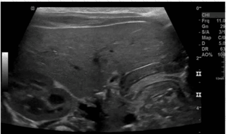
The ultrasound image clearly demonstrates a thickened and elongated pyloric muscle with a visible channel, which is characteristic of hypertrophic pyloric stenosis (HPS). This condition is most commonly seen in male infants between 2 and 8 weeks of age who present with non-bilious projectile vomiting, dehydration, and a palpable “olive-like” mass in the right upper quadrant.
Ultrasound is the imaging modality of choice and is highly sensitive and specific for diagnosing pyloric stenosis.
Key sonographic criteria for HPS:
Muscle thickness >3 mm
Pyloric channel length >15–17 mm
“Target sign” or “doughnut sign” on transverse imaging (concentric rings)
“Cervix” or “railroad track sign” on longitudinal imaging (elongated canal with echogenic center)
Differentiation from other options:
A. Intussusception: Also shows a target sign, but it occurs in the right lower quadrant or periumbilical region, not in the gastric antrum.
C. Hydronephrosis: Refers to dilation of the renal pelvis and calyces — not gastrointestinal.
D. Gastritis: May show gastric wall thickening but lacks the distinct elongated, thickened pyloric muscle seen here.
[References:, Rumack CM, Wilson SR, Charboneau JW, Levine D. Diagnostic Ultrasound. 5th Edition. Elsevier, 2018. Chapter: Gastrointestinal Tract, pp. 474–479., American College of Radiology (ACR) Appropriateness Criteria – Vomiting in Infants Up to 3 Months of Age., AIUM Practice Parameter for the Performance of a Pediatric Abdominal Ultrasound Examination, 2020., , , ]
Submit