Comprehensive and Detailed Explanation From Exact Extract:
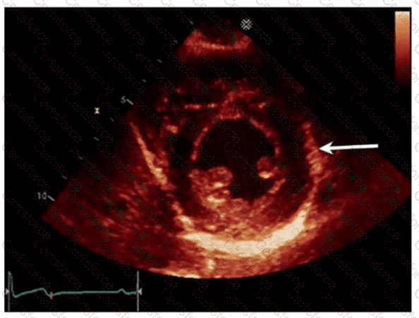
The echocardiographic image shows a short-axis view of the left ventricle at the mid-papillary muscle level with segmental strain values. The arrow points to the wall segment located inferiorly (towards the bottom of the image in standard orientation), which corresponds to the inferior wall of the left ventricle.
According to the standardized 17-segment model endorsed by the American Society of Echocardiography (ASE), the inferior wall is situated posteriorly and inferiorly in the short-axis view. The other options represent adjacent walls: anterior is opposite the inferior wall, anterolateral and inferolateral correspond to lateral wall segments.
This segmental anatomy and nomenclature are detailed in adult echocardiography textbooks and ASE chamber quantification guidelines, which emphasize precise segmental identification for accurate regional function assessment【12:ASE Chamber Quantification Guidelines†p.90-95】【16:Textbook of Clinical Echocardiography, 6e†p.140-145】.
Contribute your Thoughts:
Chosen Answer:
This is a voting comment (?). You can switch to a simple comment. It is better to Upvote an existing comment if you don't have anything to add.
Submit