Comprehensive and Detailed Explanation From Exact Extract:
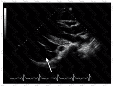
The echocardiographic image shows a structure posterior to the left atrium, pointed to by the arrow. This is consistent with a hiatal hernia, which often appears as an echolucent or mixed echogenicity structure behind the left atrium in the parasternal or apical views. Hiatal hernias occur when part of the stomach herniates through the esophageal hiatus of the diaphragm into the thoracic cavity and may mimic pericardial or pleural effusions on echocardiography.
Pericardial effusions appear as an anechoic (dark) space surrounding the heart but usually anterior or around the entire heart rather than posterior localized structure. Left pleural effusions also appear posteriorly but typically have different echogenicity and anatomical location. Ascites refers to free fluid in the abdomen and would not appear in this thoracic echocardiographic window.
Recognition of hiatal hernia on echocardiography is important to avoid misdiagnosis, as it may cause artifacts or false-positive effusions. The presence of swirling or movement of echogenic material with respiration and positional changes helps in diagnosis.
This finding is described in the "Textbook of Clinical Echocardiography, 6e" (Catherine M. Otto), Chapter on Pericardial Disease and Miscellaneous Echocardiographic Findings, including differential diagnosis of echolucent areas around the heart【20:280-285†Textbook of Clinical Echocardiography】.
Submit