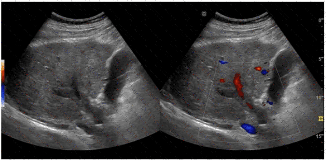
The sonographic images depict an acute thrombotic process involving the portal venous system. The absence of cavernous transformation in the setting of portal vein thrombus indicates that the process is acute. In chronic portal vein thrombosis, collateral vessels form in the porta hepatis to bypass the obstruction, a process known as cavernous transformation.
Sonographic features suggesting acute portal vein thrombosis:
Echogenic thrombus within the portal vein lumen
Absence of flow on color Doppler
Enlarged portal vein diameter early in the process
No evidence of cavernous transformation (i.e., no serpiginous collateral vessels at porta hepatis)
Cavernous transformation is a hallmark of chronic portal vein thrombosis and takes weeks to months to develop. Therefore, its absence on ultrasound supports an acute diagnosis.
Differentiation from other options:
A. Free fluid: Non-specific and may or may not be present in hepatic vascular thrombosis.
B. Ductal dilatation: Related to biliary obstruction, not portal or hepatic venous thrombosis.
C. Hepatic vein thrombosis: Seen in Budd-Chiari syndrome, which affects hepatic outflow, not portal inflow.
[References:, Rumack CM, Wilson SR, Charboneau JW, Levine D. Diagnostic Ultrasound. 5th Edition. Elsevier, 2018. Chapter: Portal Venous System, pp. 105–108., American Institute of Ultrasound in Medicine (AIUM) Practice Parameter for the Performance of Hepatic Doppler Ultrasound Examinations, 2020., Radiopaedia.org. Cavernous transformation of the portal vein: https://radiopaedia.org/articles/cavernous-transformation-of-the-portal-vein, ]
Submit