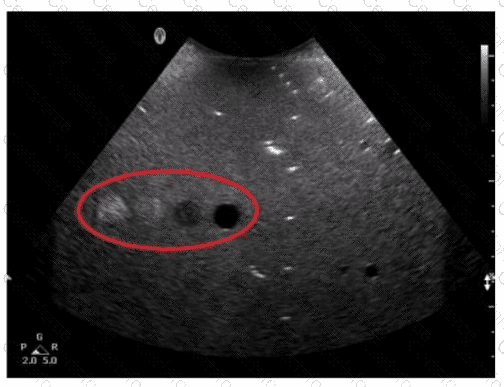
The tissue-equivalent phantom image with the red oval indicates an area where axial resolution can be evaluated. Axial resolution refers to the ability to distinguish between two structures that are close together along the axis of the ultrasound beam. It is determined by the spatial pulse length (SPL) of the ultrasound wave. In phantoms, this is typically tested by observing the ability to separate closely spaced targets along the beam's path.
[References:, ARDMS Sonography Principles & Instrumentation Guidelines, Hedrick WR, Hykes DL, Starchman DE.Ultrasound Physics and Instrumentation. 4th ed. Philadelphia, PA: Elsevier Saunders; 2005., , ]
Submit